01 // LAB CAPABILITIES

Analytical techniques for researchers and clinicians

Services are run in-house in Gifu. Contact the laboratory to discuss study design, sample requirements and turnaround before submitting.

SL-01 ● ACCEPTING SAMPLES

UV fluorescence microscopy & amyloid burden

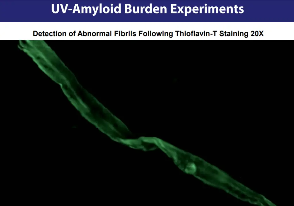
Thioflavin-T staining of dried blood smears, imaged under UV excitation to visualise and assess amyloid fibril structures.

SPECIMEN
Capillary blood smear on glass slide
STAIN
Thioflavin-T
OUTPUT
Image report + recorded video review
SL-02 ● BY ENQUIRY

RT-QuIC prion seeding assay

Real-Time Quaking-Induced Conversion amplifies misfolded prion protein and detects it through changes in fluorescence — the method used in diagnosing prion diseases such as Creutzfeldt-Jakob disease.

TARGET
Misfolded prion protein (PrPSc)
READOUT
Real-time fluorescence kinetics
SL-03 ● BY ENQUIRY

Nanopore sequencing

Long-read DNA and RNA sequencing in real time. Each nucleotide is identified by its characteristic change in ionic current as the strand passes through a membrane nanopore, with no amplification required.

INPUT
DNA or RNA
READOUT
Long-read sequence data
SL-04 ● BY ENQUIRY

Confocal Raman spectroscopy

Research-grade confocal Raman microscopy on a Renishaw inVia™ system for non-destructive chemical identification and molecular fingerprinting of materials.

SYSTEM
Renishaw inVia™ confocal Raman
READOUT
Raman spectra & spectral maps
SL-05 ● BY ENQUIRY

SEM-EDX elemental analysis

Scanning electron microscopy with energy-dispersive X-ray spectroscopy for high-magnification surface imaging and elemental composition mapping.

IMAGING
Scanning electron microscopy
READOUT
Elemental spectra & maps
SL-06 ● CONSULTANCY

Neurophysiology & deep brain stimulation

Scientific consultancy on large-scale cortico-basal ganglia network physiology and the application of deep brain stimulation to movement and neuropsychiatric disorders.

AREAS
Network physiology, DBS, disease models
FORMAT
Study design & research collaboration
02 // WORKFLOW

From specimen to report

  1. STAGE 01 →
    Intake
    Slides arrive with health summary and contact email.
  2. STAGE 02 →
    Preparation
    Specimens logged and stained for the requested technique.
  3. STAGE 03 →
    Acquisition
    Imaging, spectra or sequence data captured on-instrument.
  4. STAGE 04 →
    Analysis
    Results interpreted by the principal investigator.
  5. STAGE 05 ✓
    Report
    Confidential results by email, with optional recorded video review.
03 // INSTRUMENTATION

Research-grade imaging and spectroscopy

The laboratory's spectroscopy platform is a Renishaw inVia™ confocal Raman microscope — a research-grade system refined over more than two decades and widely used for its data quality and speed of acquisition.

It is complemented by UV fluorescence microscopy for stained specimens, scanning electron microscopy with EDX elemental analysis, nanopore sequencing and RT-QuIC protein-misfolding assays.

EQUIPMENT_SUMMARY
PLATFORM APPLICATION
Renishaw inVia™ confocal RamanChemical identification, spectral mapping
UV fluorescence microscopeThioflavin-T amyloid imaging
SEM with EDX detectorSurface imaging, elemental analysis
Nanopore sequencerLong-read DNA / RNA sequencing
RT-QuIC plate readerPrion seeding activity
Thioflavin-T stained fibrils fluorescing green under UV light ThT · UV ex · 20×
FIG.02 — THIOFLAVIN-T AMYLOID FLUORESCENCESL-01
04 // CURRENT RESEARCH · PRJ-PRION-01

Prion Research Investigation Project

STATUS: OPEN TO RESEARCH COLLABORATION

An investigation at the intersection of virology, immunology and neurodegenerative disease, testing whether SARS-CoV-2 proteins — particularly the spike protein — or mRNA vaccine components show prion-like misfolding activity, and whether any such activity carries long-term neurological risk.

Prions are misfolded proteins that induce normal proteins to misfold, causing progressive and fatal neurodegenerative conditions such as Creutzfeldt-Jakob disease. The project aims to answer the question with evidence from rigorous laboratory work.

RESEARCH_QUESTIONS

  1. Q1Does SARS-CoV-2 contain prion-like domains capable of misfolding?
  2. Q2Can infection, or spike protein exposure via mRNA vaccination, trigger prion formation in cells or animal models?
  3. Q3What molecular mechanisms of misfolding and aggregation would be involved?
  4. Q4What are the long-term implications for neurological health and public health policy?
  • IN VITRO
    Human and animal cell cultures to assess misfolding and aggregation.
  • IN VIVO
    Transgenic mouse models of prion formation and neurodegeneration.
  • IN SILICO
    Computational folding models to predict prion-like motifs.
  • PMCA · RT-QuIC
    Biochemical detection of prion-like seeding activity.
  • NEUROPATHOLOGY
    Post-mortem analysis for prion-related neurodegeneration.
Illustration of brain network science
05 // PRINCIPAL INVESTIGATOR

Dr Kevin W. McCairn, PhD

Chief Executive Officer · Systems Neuroscientist

Dr McCairn has published extensively on large-scale network interactions of the cortico-basal ganglia circuits. He developed the first primate model of Tourette syndrome, and has worked on the efficient use of deep brain stimulation for both movement and neuropsychiatric disorders.

NEUROBIOLOGY NEUROPHYSIOLOGY NEURODEGENERATIVE DISEASE BRAIN STIMULATION BRAIN PHYSIOLOGY
06 // SAMPLE SUBMISSION · PROTOCOL SUB-UVA-01

Blood smear collection for UV microscopy & amyloid burden

Follow each step below. All submissions are handled confidentially. Fees are invoiced by email once analysis is complete.

Watch: how to take & send a blood sample
VIDEO · SAMPLE COLLECTION METHODOLOGY · OPENS IN A NEW TAB
  1. 01

    Gather materials

    • Pre-cleaned glass microscope slides — no coverslips
    • Single-use safety lancet
    • Sterile alcohol prep swab
    • Microscope slide mailer
  2. 02

    Prepare the smear

    Clean the fingertip with the swab, collect a drop of blood with the lancet and prepare a smear on the slide. Allow it to air-dry fully. A methodology video is available on request.

  3. 03

    Include your details

    Enclose a brief health summary (for example diabetes or long COVID), your name, return address and the email address where results should be sent.

  4. 04

    Pack & ship

    Place dried slides in a grooved slide box or mailer, cushioned inside a sturdy outer box. Ship by post or carrier at room temperature — do not freeze.

SHIP_TO
Dr Kevin McCairn
Synaptek
Kani, Nishi Katabira 238-1
509-0265 Japan
RESULTS

Findings are sent to your email with an optional recorded video review by Dr McCairn. Please check your spam folder if results do not arrive.

Analyses are performed for research purposes and are not a medical diagnosis. Discuss any findings with your healthcare provider.